OSF Innovation Brings Beating Hearts to Life in VR for Safer Surgeries
Peoria, IL – September 2025 — Surgeons can now view a patient’s heart beating in virtual reality thanks to a breakthrough technology developed by OSF Innovation in collaboration with Bradley University and the University of Illinois Urbana-Champaign. The new 4D Heart platform gives clinicians a dynamic, patient-specific digital twin of the heart in motion, improving surgical planning and potentially reducing risk during complex procedures.
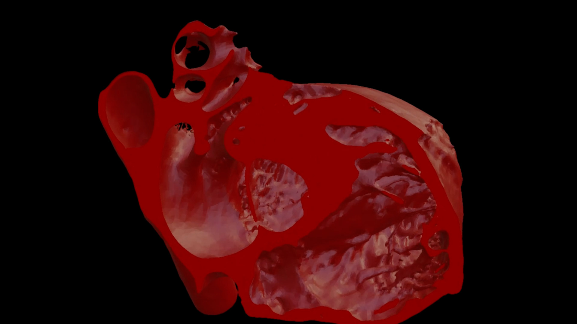
Traditional 2D CT images provide static snapshots, forcing surgeons to mentally piece together a moving organ.
The 4D Heart changes this by rendering every phase of a patient’s cardiac cycle, enabling doctors to see how the heart twists and contracts in real time.
“It’s like a digital flipbook of the heart,” said Dr. Matthew Bramlet, Director of the Advanced Imaging and Modeling Lab at OSF Innovation. “Surgeons can interact with these models in ways they’ve never been able to before.”
AI Makes It Practical for Real-World Use
Initially, creating a 4D Heart model took an entire summer. By late 2024, engineers trained advanced neural networks on hundreds of high-quality CT scans, fully automating the process. What once required months now happens with a single click, making the technology viable for widespread clinical use.
The AI engine even identified structures like heart valves—historically difficult to capture with CT scans—offering new insights for procedures such as valve replacements and cases of hypertrophic obstructive cardiomyopathy.
Transforming Surgical Decision-Making
Hospitals across the U.S. are now sending scans to OSF for conversion into 4D files, even if they lack the hardware to view them yet. The technology has already impacted surgical planning:
- Confirming strategies for certain procedures
- Changing approaches in complex cases, including pediatric heart surgeries
Dr. Bramlet predicts the technology will soon become standard of care, allowing surgeons to move from planning with static images to fully immersive, dynamic models.
Looking Ahead
The ultimate goal is a seamless workflow where a patient’s CT or MRI scan automatically generates a digital twin heart, complete with AI-driven analytics like ejection fraction or blood flow strain. While adoption depends on health system investment and reimbursement policies, research funding—such as from the Jump ARCHES endowment—is driving current progress.
“Seeing is believing,” Dr. Bramlet said. “This is the future of surgical planning and patient care.”